Digital dental X-rays, including intraoral cameras and panoramic scans, have transformed oral health assessments by enabling dentists to detect caries, gum disease, and impactions more accurately. They offer benefits like instant high-res images, electronic storage, reduced radiation exposure (80% less than film methods), and minimized handling time. Despite higher initial costs and interpretation requirements, advancements make digital X-rays more accessible and crucial for modern dentistry, particularly in diagnosing and treating conditions requiring precise evaluations, such as fillings or crowns. High-quality image clarity and standardized patient positioning ensure accurate diagnosis and successful treatments like dental implants.
Digital dental X-rays have revolutionized oral imaging, offering improved efficiency and precision compared to traditional film methods. This article explores the various types of digital dental X-rays, their practical applications, and the advantages they bring in terms of image quality and patient safety. We’ll delve into quality assurance measures and techniques for interpreting digital X-ray images accurately. Discover how these advanced technologies are shaping modern dental practices and enhancing diagnostic capabilities with enhanced resolution and reduced radiation exposure.
- Types of Digital Dental X-Rays and Their Uses
- Advantages and Disadvantages Over Traditional Film X-Rays
- Quality Assurance and Image Interpretation Techniques
Types of Digital Dental X-Rays and Their Uses
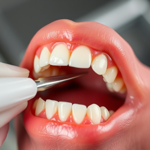
Digital dental X-rays have transformed the way dental professionals assess oral health and perform various procedures. These advanced imaging tools offer several types of digital radiography, each with specific applications. One common form is the intraoral camera, a small, wireless device that captures high-resolution images of teeth and gums from within the mouth. This technology allows dentists to detect caries, gum disease, or other abnormalities not visible during a visual examination.
Another type, digital panoramic X-rays, provides a wide view of the entire dentition, including jawbones and surrounding structures. They are invaluable for identifying impactions, assessing oral development in children, and planning orthodontic treatments. In restorative dentistry, digital dental X-rays play a crucial role in diagnosing tooth decay and determining the extent of damage, aiding in decisions regarding fillings, crowns, or dental bonding to repair and restore teeth effectively.
Advantages and Disadvantages Over Traditional Film X-Rays
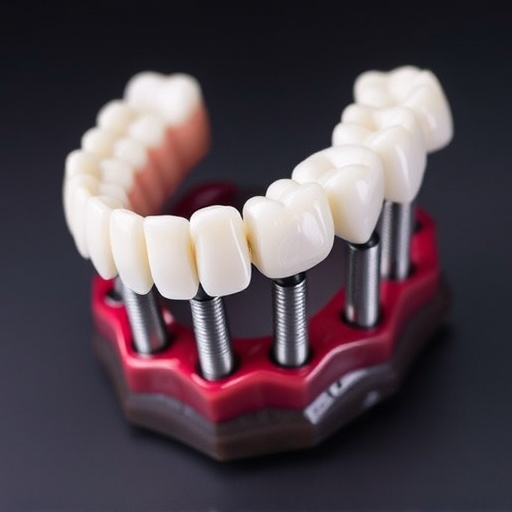
Digital dental X-rays offer several advantages over traditional film X-rays. They provide instant, high-resolution images that can be easily stored and shared electronically, eliminating the need for physical films to be developed and stored. This not only reduces handling time but also minimizes potential errors in image transfer or loss of files. Additionally, digital X-rays reduce patient exposure to radiation by up to 80% compared to film methods, making them a safer option for regular dental check-ups, especially in children’s dentistry.
Despite these benefits, there are some disadvantages to consider. Digital sensors and imaging equipment can be more expensive upfront than traditional film systems. Moreover, while digital X-rays offer clearer images, they may require specialized knowledge to interpret accurately, which could pose challenges for less experienced dental professionals. Nevertheless, with advancements in technology, the accessibility and affordability of digital dental X-rays continue to improve, making them a game-changer in modern dentistry, particularly when it comes to diagnosing conditions that require precise evaluations, such as identifying the need for dental fillings or crafting intricate dental crowns.
Quality Assurance and Image Interpretation Techniques
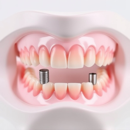
Ensuring high-quality digital dental X-rays is paramount for accurate diagnosis and comprehensive dental care. Modern technology offers a range of quality assurance techniques to optimize image interpretation. One key method involves using specialized software that enhances image clarity, allowing dentists to detect even minute anomalies. This process includes adjusting contrast, brightness, and resolution, ensuring every detail is visible.
Additionally, proper patient positioning and consistent technique are vital for maintaining consistency in X-ray images. Dentists employ standardized protocols during routine oral exams and check-ups to minimize errors and ensure reliable results. These practices form the backbone of effective digital dental X-ray interpretation, facilitating the early detection of caries, gum diseases, and other conditions, ultimately supporting the successful implementation of treatments such as dental implants.
Digital dental X-rays have revolutionized oral imaging, offering improved quality, reduced exposure to radiation, and enhanced efficiency. By understanding the types of digital X-ray techniques, their advantages over film, and implementing strict quality assurance protocols for image interpretation, healthcare providers can ensure optimal patient care using this cutting-edge technology. The future of dental diagnostics looks bright with continued advancements in digital dental x-rays.


